44 sarcomere diagram labeled
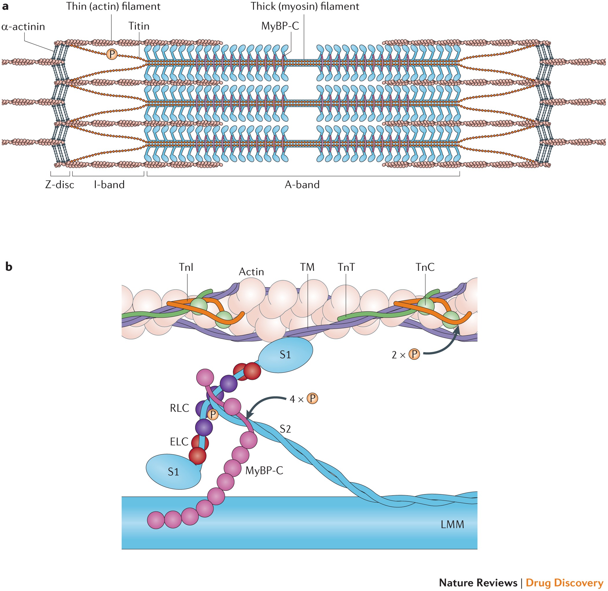
Label the Sarcomere Structure Diagram | Quizlet Start studying Label the Sarcomere Structure. Learn vocabulary, terms, and more with flashcards, games, and other study tools. Anatomy of the cardiac sarcomere. (A) Diagram of the basic organization ... The sarcomere forms the basic contractile unit in the cardiomyocytes of the heart. Thin filaments composed of actin are anchored at the Z line and form transient sliding interactions with thick ...
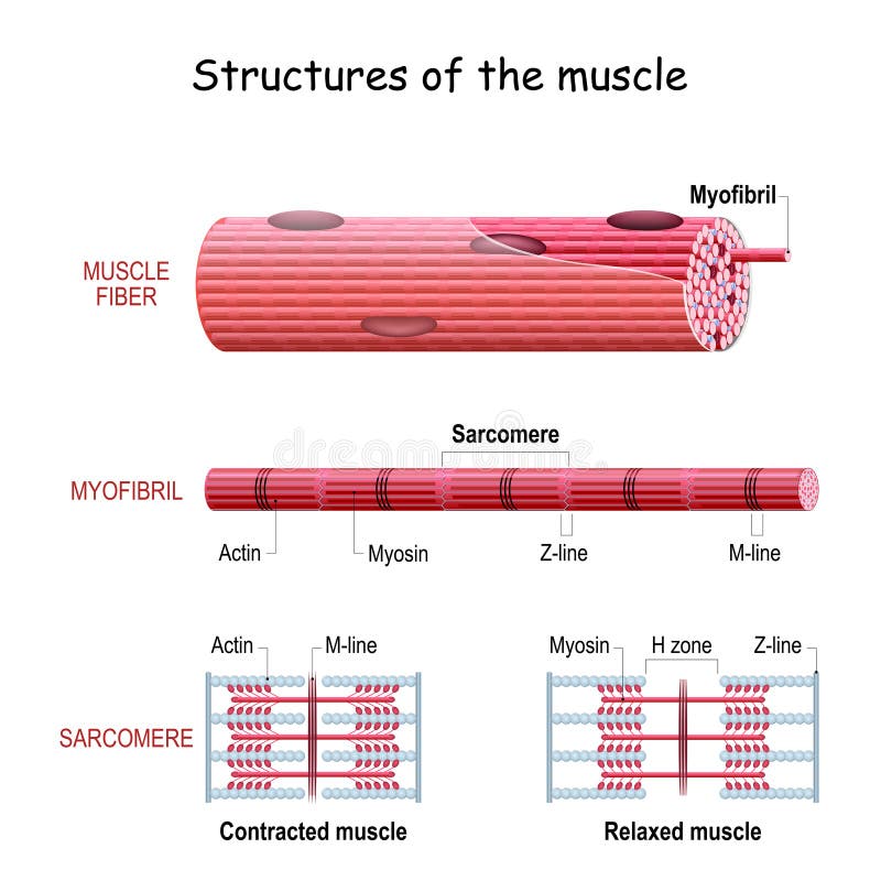
Comparison of a relaxed and contracted sarcomere (A) The basic organization of a sarcomere subregion, showing the centralized location of myosin (A band). Actin and the z discs are shown in red. (B) A conceptual diagram representing the ...

Sarcomere diagram labeled
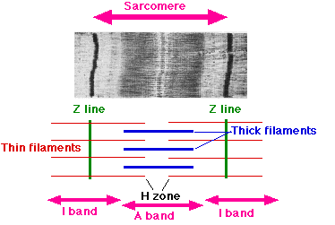
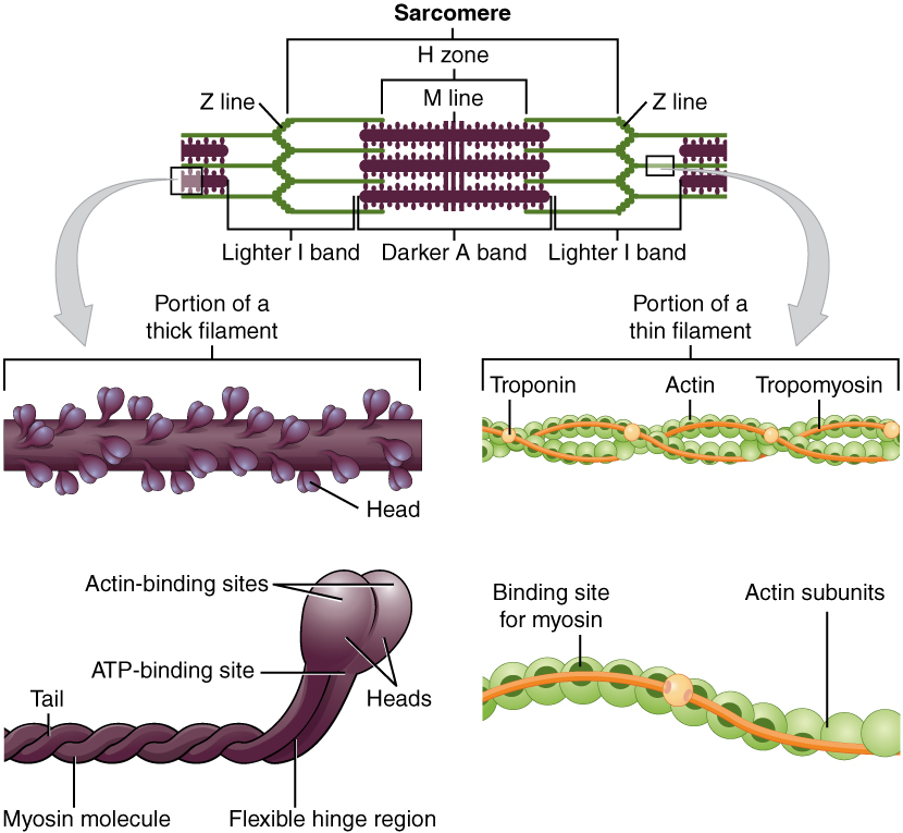
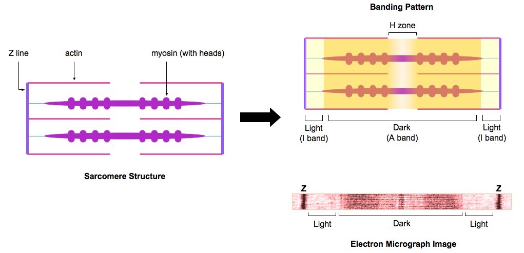
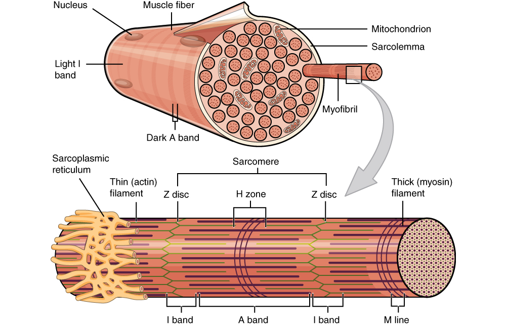
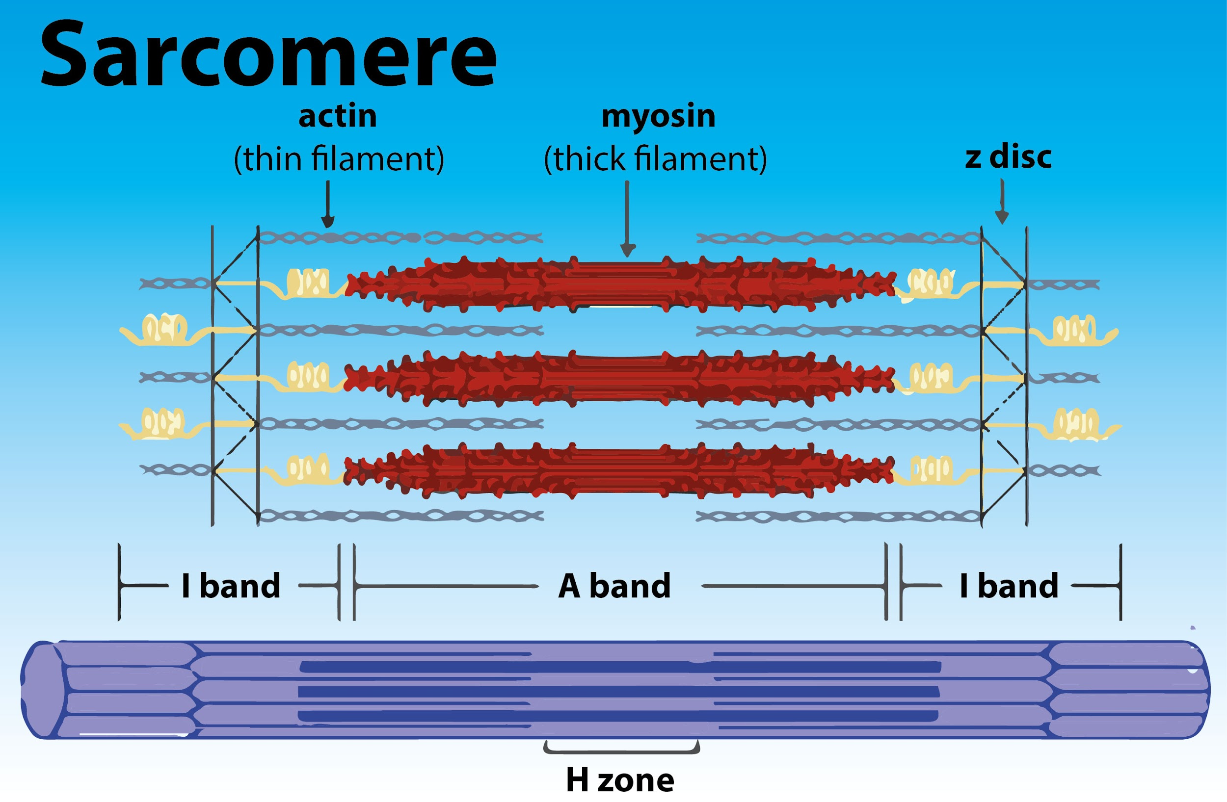
quizlet.com › 388081861 › sarcomere-labeled-diagramsarcomere labeled diagram Diagram | Quizlet sarcomere labeled diagram + − Flashcards Learn Test Match Created by ruthiewilhelm Terms in this set (8) i band light band m line middle of sarcomere myosin thick filament a band dark band h zone only myosin actin thin filaments z line end of sarcomere cross bridges when the myosin heads interact with thin filaments during a contraction MGreer5 quizlet.com › 245378977 › sarcomere-labeling-diagramSarcomere Labeling Diagram | Quizlet Sarcomere The smallest contractile unit of muscle; extends from one Z disc to the next H Band The band at the middle of the A Band, where only myosin is found A Band The darkest area that runs the length of the myosin, including where actin and myosin overlap I Band On either side of the A Band is the I band, where only the Actin is found Z Disk › scitable › topicpageSliding Filament Theory, Sarcomere, Muscle Contraction, Myosin |... (B) A conceptual diagram representing the connectivity of molecules within a sarcomere. A person standing between two bookcases (z bands) pulls them in via ropes (actin). Myosin (M) is analogous ...
Sarcomere diagram labeled. Sarcomere (Muscle) Coloring - The Biology Corner The enter muscle fiber is surrounded by the sarcolemma (D), color this membrane brown. If expanded, the light and dark bands are shown as individual thick and thin filaments. Color the thick filaments (not labeled) red and the thin filaments blue. The Z line is the boundary between sarcomeres, named after its shape. Color the Z-line orange. anatomy of a sarcomere Sarcomere Diagram Labeled schematron.org. sarcomere labeled filaments. Sliding Filament Sarcomere - YouTube . sarcomere filament sliding anatomy projects school. Contracted State Of Sarcomere In 2021 | Muscle, Muscle Contraction . ibiologia.com › sarcomereSarcomere | Definition, Structure, & Sliding Filament Theory Aug 10, 2019 · Sarcomere Model: The functional sarcomere model describes briefly the sliding filament theory of skeletal muscle contraction in an understandable manner. Configuration with including complete sarcomere, and function of thin filaments (having three proteins; actin, troponin, and tropomyosin) and thick filaments. The M-line and Z-line are identifiable. thebiologynotes.com › sarcomereSarcomere- Definition, Structure, Diagram, and Functions Jul 7, 2022 · A sarcomere is a complex multicomponent biological system and functional unit of striated muscle which plays a vital role in transforming the chemical energy released upon the ATP hydrolysis into mechanical work. Skeletal muscles are made up of the basic unit called a sarcomere and all voluntary movement is initiated by this skeletal muscle.
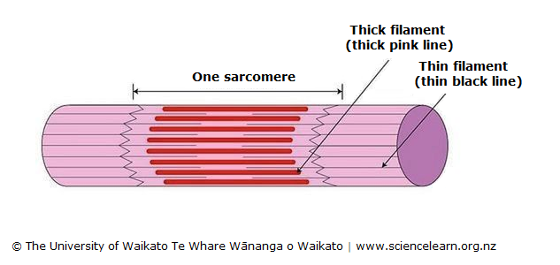
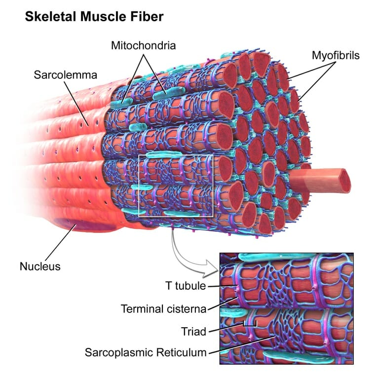
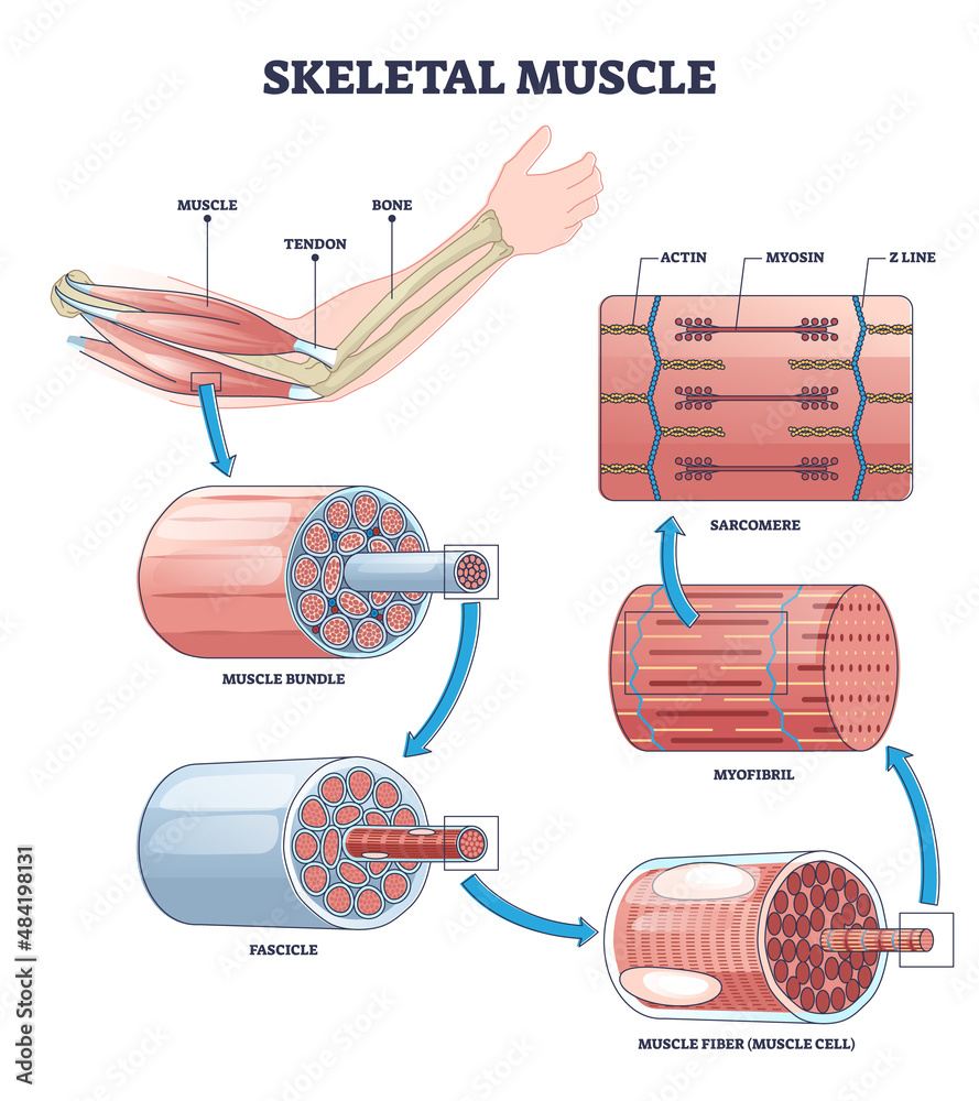
Sliding Filament Model of Contraction | Biology for Majors II The mechanism of contraction is the binding of myosin to actin, forming cross-bridges that generate filament movement (Figure 1). Figure 1. When (a) a sarcomere (b) contracts, the Z lines move closer together and the I band gets smaller. The A band stays the same width and, at full contraction, the thin filaments overlap. Sarcomere: anatomy, structure and function | Kenhub The sarcomere is the main contractile unit of muscle fiber in the skeletal muscle.Each sarcomere is composed of protein filaments (myofilaments) that include mainly the thick filaments called myosin, and thin filaments called actin.The bundles of myofilaments are called myofibrils.. The structure of the sarcomere is traditionally described with dark and light bands visible under the microscope. Sarcomere - Definition, Structure, Function and Quiz - Biology Dictionary A sarcomere is the functional unit of striated muscle. This means it is the most basic unit that makes up our skeletal muscle. Skeletal muscle is the muscle type that initiates all of our voluntary movement. Herein lies the sarcomere's main purpose. Sarcomeres are able to initiate large, sweeping movement by contracting in unison. Diagram Of Sarcomere The sarcomere is the contractile unit of muscle. This means it is the part of muscle Diagram of the Sarcomere. Source: . A dark stripe called a Z disc marks the ends of one sarcomere and the beginning Look at the diagram above and realize what happens as a muscle contracts.Mass Haul Diagram Explained. Whirlpool Duet Dryer Parts Diagram.
diagramweb.net › labeled-sarcomere-diagramLabeled Sarcomere Diagram Jan 23, 2019 · A sarcomere is the basic unit of striated muscle tissue. It is the repeating unit between two Z lines. Skeletal muscles are composed of tubular muscle cells which. Sarcomeres are composed of thick filaments and thin filaments. The thin filaments Look at the diagram above and realize what happens as a muscle contracts. Sarcomere - an overview | ScienceDirect Topics A sarcomere is the basic contractile unit of muscle fiber. Each sarcomere is composed of two main protein filaments—actin and myosin—which are the active structures responsible for muscular contraction. ... Figure 2-6 diagrams the cytoskeleton of the sarcomere and its relationship to the contractile proteins. 6 The M line and the Z disc ... anatomy of sarcomere - Microsoft Labeled Sarcomere Diagram schematron.org. sarcomere labeled diagram muscle unit skeletal striated tissue basic. PPT - Chapter 9 PowerPoint Presentation, Free Download - ID:433984 . sarcomere structure muscle band ppt organization powerpoint presentation figure 4a chapter zone line slideserve. Sarcomere Structure | Anatomy And ... anatomy of sarcomere hand labeled bones diagram anatomy human label bone body thumb part labels pad zazzle board poster outline press arthritic changes. PPT - Figure 10.1 The Organization Of Skeletal Muscles PowerPoint . figure sarcomere structure skeletal part muscles organization. Sarcomere Anatomy Quiz . sarcomere quiz ...
› scitable › topicpageSliding Filament Theory, Sarcomere, Muscle Contraction, Myosin |... (B) A conceptual diagram representing the connectivity of molecules within a sarcomere. A person standing between two bookcases (z bands) pulls them in via ropes (actin). Myosin (M) is analogous ...
quizlet.com › 245378977 › sarcomere-labeling-diagramSarcomere Labeling Diagram | Quizlet Sarcomere The smallest contractile unit of muscle; extends from one Z disc to the next H Band The band at the middle of the A Band, where only myosin is found A Band The darkest area that runs the length of the myosin, including where actin and myosin overlap I Band On either side of the A Band is the I band, where only the Actin is found Z Disk
quizlet.com › 388081861 › sarcomere-labeled-diagramsarcomere labeled diagram Diagram | Quizlet sarcomere labeled diagram + − Flashcards Learn Test Match Created by ruthiewilhelm Terms in this set (8) i band light band m line middle of sarcomere myosin thick filament a band dark band h zone only myosin actin thin filaments z line end of sarcomere cross bridges when the myosin heads interact with thin filaments during a contraction MGreer5












Komentar
Posting Komentar