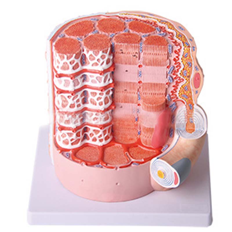
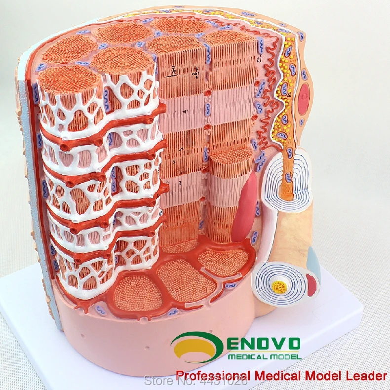
38 skeletal muscle fiber model labeled
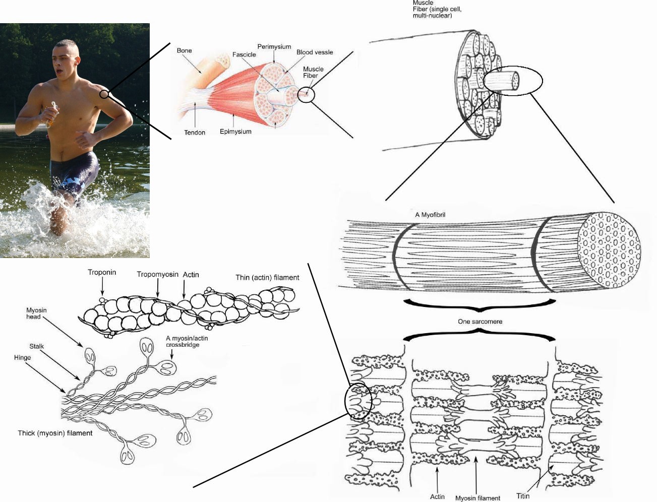
Skeletal Muscle: What Is It, Function, Location & Anatomy Skeletal muscles comprise 30 to 40% of your total body mass. They're the muscles that connect to your bones and allow you to perform a wide range of movements and functions. Skeletal muscles are voluntary, meaning you control how and when they work. Appointments 216.444.2606. Appointments & Locations. M4 – Muscle Fiber 3B – B60 Muscle Microanatomy. Page 1 of 2 ... NOTE: This is one muscle cell (muscle fiber) NOT an entire “named” skeletal muscle such as the biceps brachii.
Andrew File System Retirement - Technology at MSU WebAndrew File System (AFS) ended service on January 1, 2021. AFS was a file system and sharing platform that allowed users to access and distribute stored content. AFS was available at afs.msu.edu an…
Skeletal muscle fiber model labeled
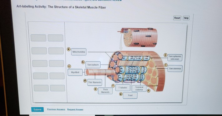
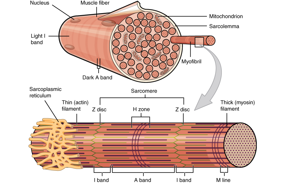
Correctly Label The Following Parts Of A Skeletal Muscle Fiber A skeletal muscle fiber is surrounded by a plasma membrane, which contains the sarcoplasm of the muscle cell. It is composed of many fibrils. These fibrils give the muscle fiber its striated appearance. The myofilaments are arranged sequentially in the sarcomere, which gives the fiber its striated look. muscle fiber model labeling Diagram | Quizlet Only $35.99/year muscle fiber model labeling How do you want to study today? Flashcards Review terms and definitions Learn Focus your studying with a path Test Take a practice test Match Get faster at matching terms Created by crlavenuePLUS Terms in this set (7) transverse tubule sarcoplasmic reticulum triad sarcolemma myofibril tech.msu.edu › about › guidelines-policiesAndrew File System Retirement - Technology at MSU Andrew File System (AFS) ended service on January 1, 2021. AFS was a file system and sharing platform that allowed users to access and distribute stored content. AFS was available at afs.msu.edu an…
Skeletal muscle fiber model labeled. Myofibril - Wikipedia WebA myofibril (also known as a muscle fibril or sarcostyle) is a basic rod-like organelle of a muscle cell. Skeletal muscles are composed of long, tubular cells known as muscle fibers, and these cells contain many chains of myofibrils. Each myofibril has a diameter of 1–2 micrometres. They are created during embryonic development in a process known as … Skeletal Muscle Fiber Types - BIO 264 Anatomy & Physiology I SKELETAL MUSCLE ORGANIZATION 7.2.1. Gross and Microscopic Structure 7.3. NEUROMUSCULAR JUNCTION, EXCITATION-CONTRACTION COUPLING, SLIDING FILAMENT THEORY, CONTRACTURES AND CRAMPS 7.3.1. Neuromuscular Junction, Excitation-Contraction Coupling, and Sliding Filament Theory 7.3.2. Muscle Contractures and Cramps 7.4. WHOLE MUSCLE CONTRACTION 7.4.1. Skeletal muscle fiber model Flashcards | Quizlet Skeletal muscle fiber model STUDY Flashcards Learn Write Spell Test PLAY Match Gravity myofibril Click card to see definition 👆 ... Click again to see term 👆 1/35 Created by yelhsamasx3 Terms in this set (35) myofibril sarcolemma 14 sarcomere 10 I (light) band 11 A (dark) band 9 transverse tubules 15 triad 16 sarcoplasmic reticulum 17 anatomy of skeletal fiber . skeletal stimulate neuron neurons fibers physiology stimulation etext. Muscle skeletal anatomy nucleus fiber label human info guws medical. Print exercise 14: microscopic anatomy and organization of skeletal. 9.4 motor neurons stimulate skeletal muscle fibers to contract: human.
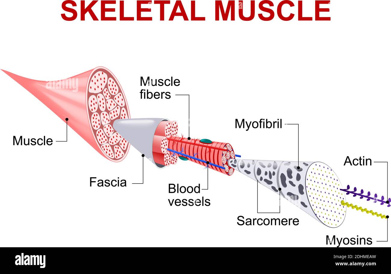
Skeletal Muscle Fiber - GetBodySmart A sarcomere is the smallest functional unit of skeletal muscle tissue, and each sarcomere has thick and thin filaments primarily composed of the proteins myosin and actin. The interaction of actin and myosin causes muscle contraction and therefore movement. Learn the physiology of skeletal muscles with the interactive tutorials and diagrams below. General Anatomy of Skeletal Muscle Fibers | GetBodySmart General Anatomy of Skeletal Muscle Fibers. Start Quiz. Identify and solve gaps in your knowledge using these interactive, spaced repetition-inspired anatomy quizzes. Learn anatomy faster and. remember everything you learn. Muscle Fiber Anatomy Strengthening lower trapezius. Muscle Fiber Anatomy. Structure of Muscle Fibers (IB Biology) - YouTube we have 9 Pics about Structure of Muscle Fibers (IB Biology) - YouTube like Skeletal Muscle Fiber, Cold Laser on Bicep Tendinitis Treatment and Shoulder Sprains and also MUSCULAR SYSTEM ANATOMY:Muscle fiber with neuromuscular junction model. Skeletal Muscle Histology Slide Identification and Labeled Diagram ... Please try to find out these structures from the skeletal muscle slide labeled images. #1. Longitudinal section of skeletal muscle #2. Cross-section of skeletal muscle #3. Skeletal muscle fibers of the longitudinal section #3. The nucleus of skeletal muscle fibers in longitudinal and cross-section #4. Cross striations of skeletal muscles #5.
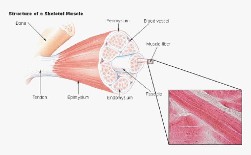
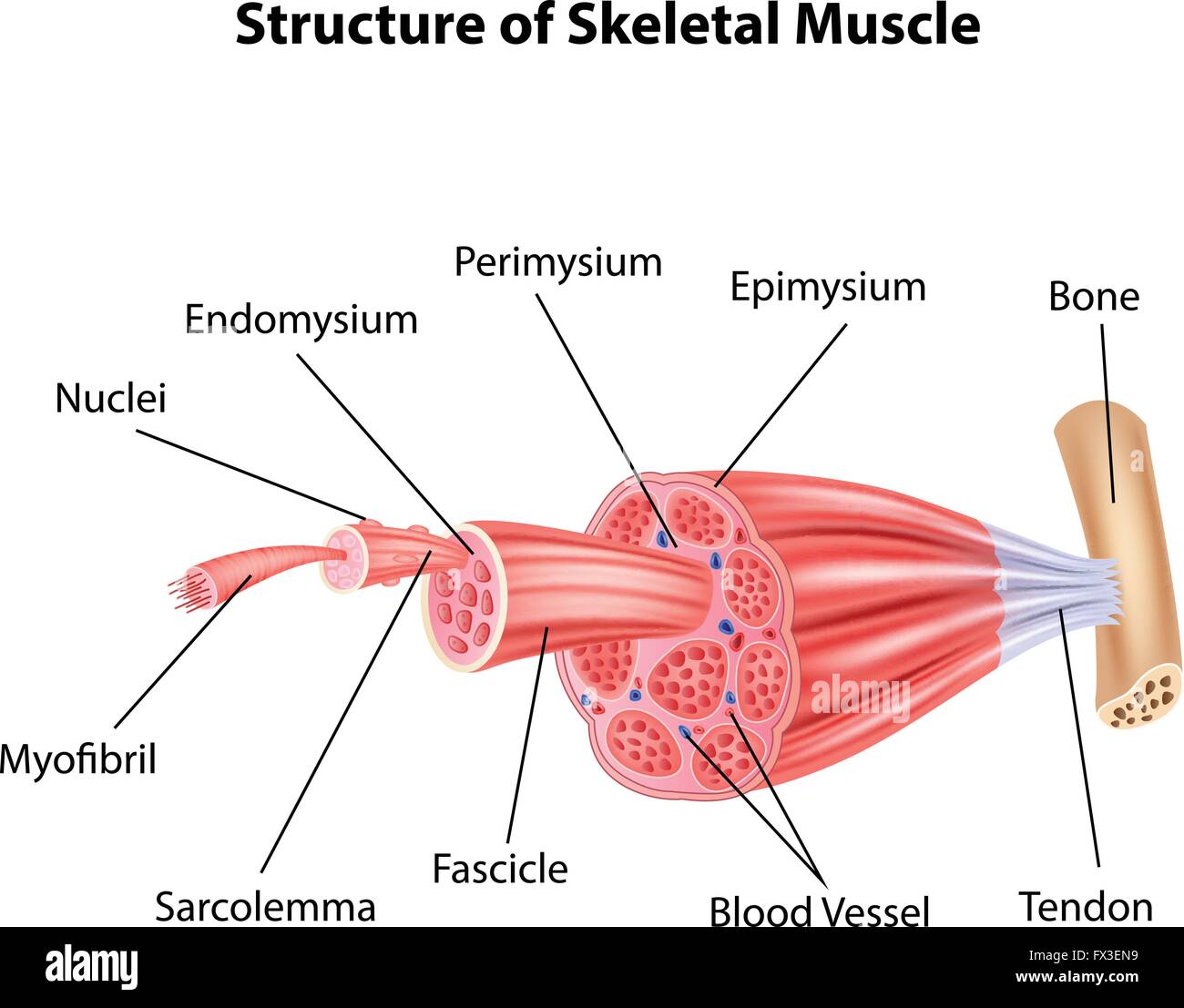
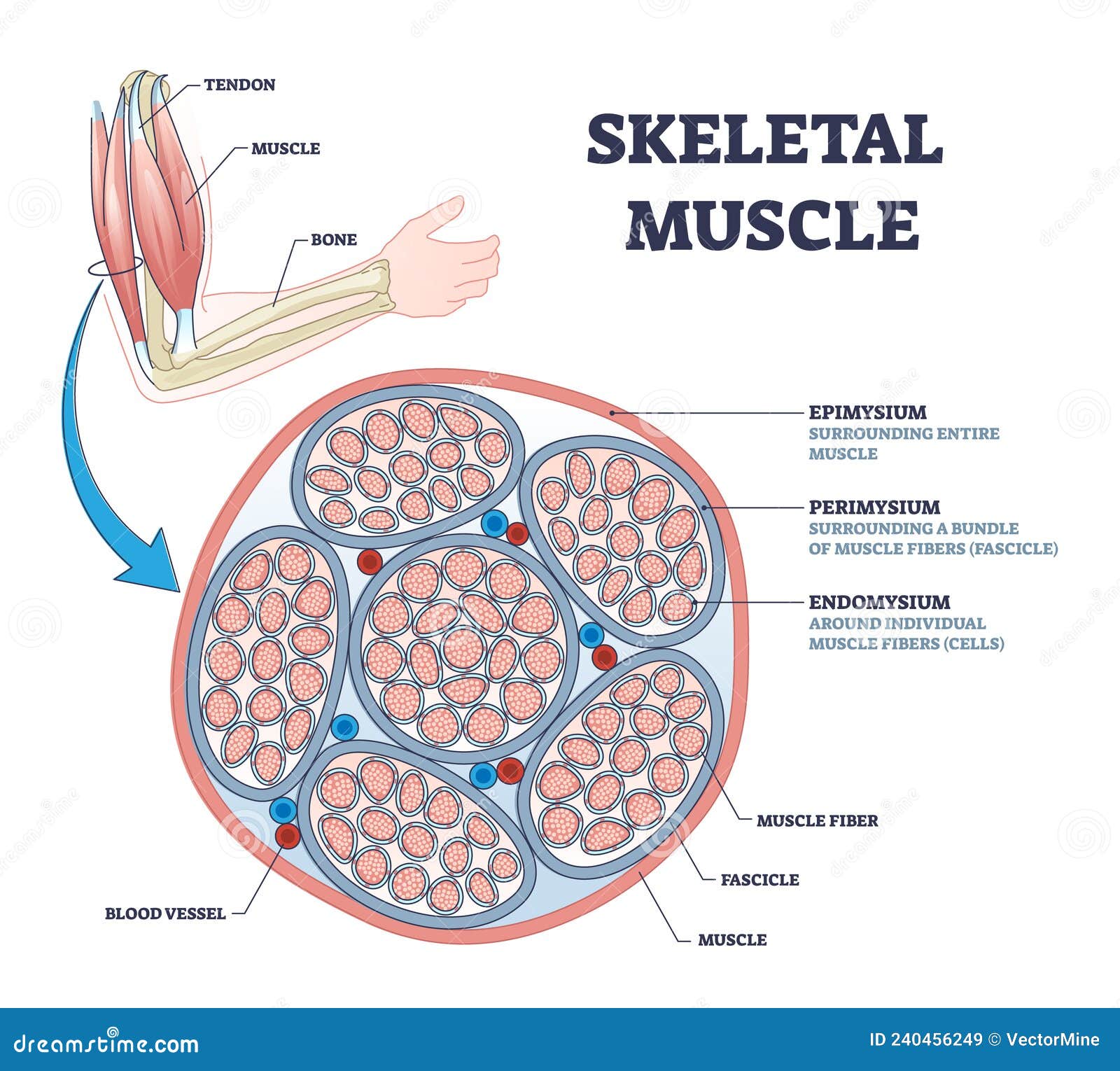
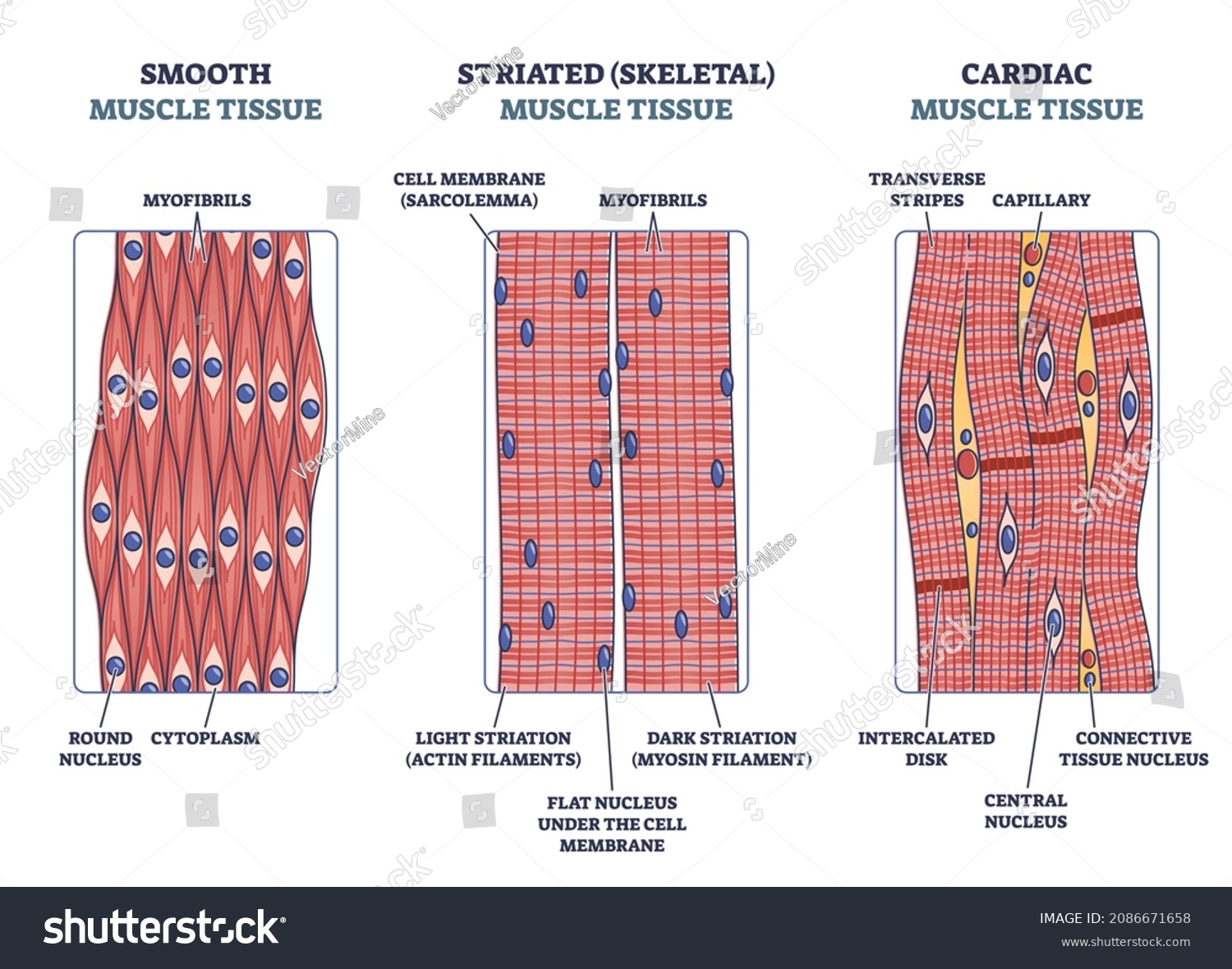
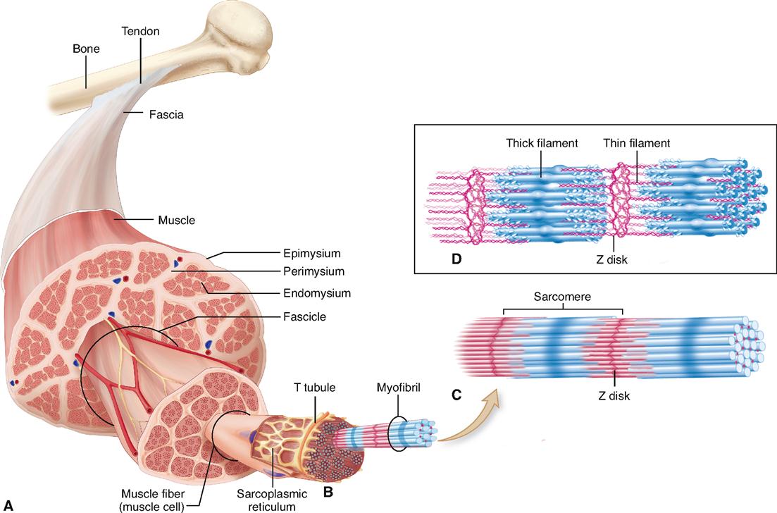
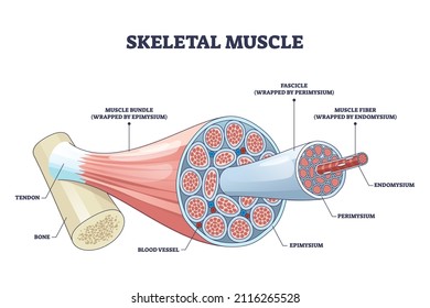
Structure of Skeletal Muscle | SEER Training Within the fasciculus, each individual muscle cell, called a muscle fiber, is surrounded by connective tissue called the endomysium. Skeletal muscle cells (fibers), like other body cells, are soft and fragile. The connective tissue covering furnish support and protection for the delicate cells and allow them to withstand the forces of contraction. Home Page: Metabolism - Clinical and Experimental Web08/09/2020 · Dear Friends and Colleagues, As Editor-in-Chief of Metabolism: Clinical and Experimental, I'm happy to share great news about the journal. Our Impact Factor has been continuously increasing over the past eleven years that I have been serving at the helm, and is now at 13.934, placing the journal amongst the top 4% of endocrinology, diabetes, and … Ultrastructure of Muscle - Skeletal - Sliding Filament - TeachMeAnatomy Ultrastructure of Muscle Cells. Muscle tissue has a unique histological appearance which enables it to carry out its function. There are three main types of muscle: Skeletal - striated muscle that is under voluntary control from the somatic nervous system. Identifying features are cylindrical cells and multiple peripheral nuclei. Skeletal muscle fiber model - Printable - PurposeGames.com About this Worksheet. This is a free printable worksheet in PDF format and holds a printable version of the quiz Skeletal muscle fiber model. By printing out this quiz and taking it with pen and paper creates for a good variation to only playing it online. This printable worksheet of Skeletal muscle fiber model is tagged. Click on the tags ...
Muscle Fibers: Anatomy, Function, and More - Healthline Skeletal muscle fibers are classified into two types: type 1 and type 2. Type 2 is further broken down into subtypes. Type 1. These fibers utilize oxygen to generate energy for movement. Type 1...
› 2073/4409/11-19 › 29872-Deoxy-D-glucose Alleviates Cancer Cachexia-Induced Muscle ... Sep 25, 2022 · Cachexia is characterized by progressive weight loss accompanied by the loss of specific skeletal muscle and adipose tissue. Increased lactate production, either due to the Warburg effect from tumors or accelerated glycolysis effects from cachectic muscle, is the most dangerous factor for cancer cachexia. This study aimed to explore the efficiency of 2-deoxy-D-glucose (2-DG) in blocking Cori ...
Human Skeletal Muscle Fiber Type Classifications Muscle Fiber Typing. Initially, whole muscles were classified as being fast or slow based on speeds of shortening. 3 This division also corresponded to a morphological difference, with the fast muscles appearing white in some species, notably birds, and the slow muscles appearing red. The redness is the result of high amounts of myoglobin and a high capillary content. 3 The greater myoglobin ...
Types of Skeletal Muscle Fibers | Study.com All skeletal muscle fibers are striated with a regular arrangement of actin (thin) and myosin (thick) filaments. Skeletal muscle fibers are further defined based on whether they are slow-twitch...
Fibers of the skeletal muscle | Anatomy snippets The percentage of each muscle fiber present in an individual is determined by three factors: genetics, hormone levels within the blood, and the level of training undertaken. The skeletal muscle is one of 14 microanatomy models on the Complete Anatomy platform. Experience the minute detail of the human body in stunning 3D.
Skeletal Muscle | Anatomy and Physiology | | Course Hero Because skeletal muscle cells are long and cylindrical, they are commonly referred to as muscle fibers. Skeletal muscle fibers can be quite large for human cells, with diameters up to 100 μm and lengths up to 30 cm (7.6 in) in the Sartorius of the upper leg.During early development, embryonic myoblasts, each with its own nucleus, fuse with up to hundreds of other myoblasts to form the ...
Page: Metabolism - Clinical and Experimental Sep 08, 2020 · Dear Friends and Colleagues, As Editor-in-Chief of Metabolism: Clinical and Experimental, I'm happy to share great news about the journal. Our Impact Factor has been continuously increasing over the past eleven years that I have been serving at the helm, and is now at 13.934, placing the journal amongst the top 4% of endocrinology, diabetes, and metabolism journals as indexed in Journal ...
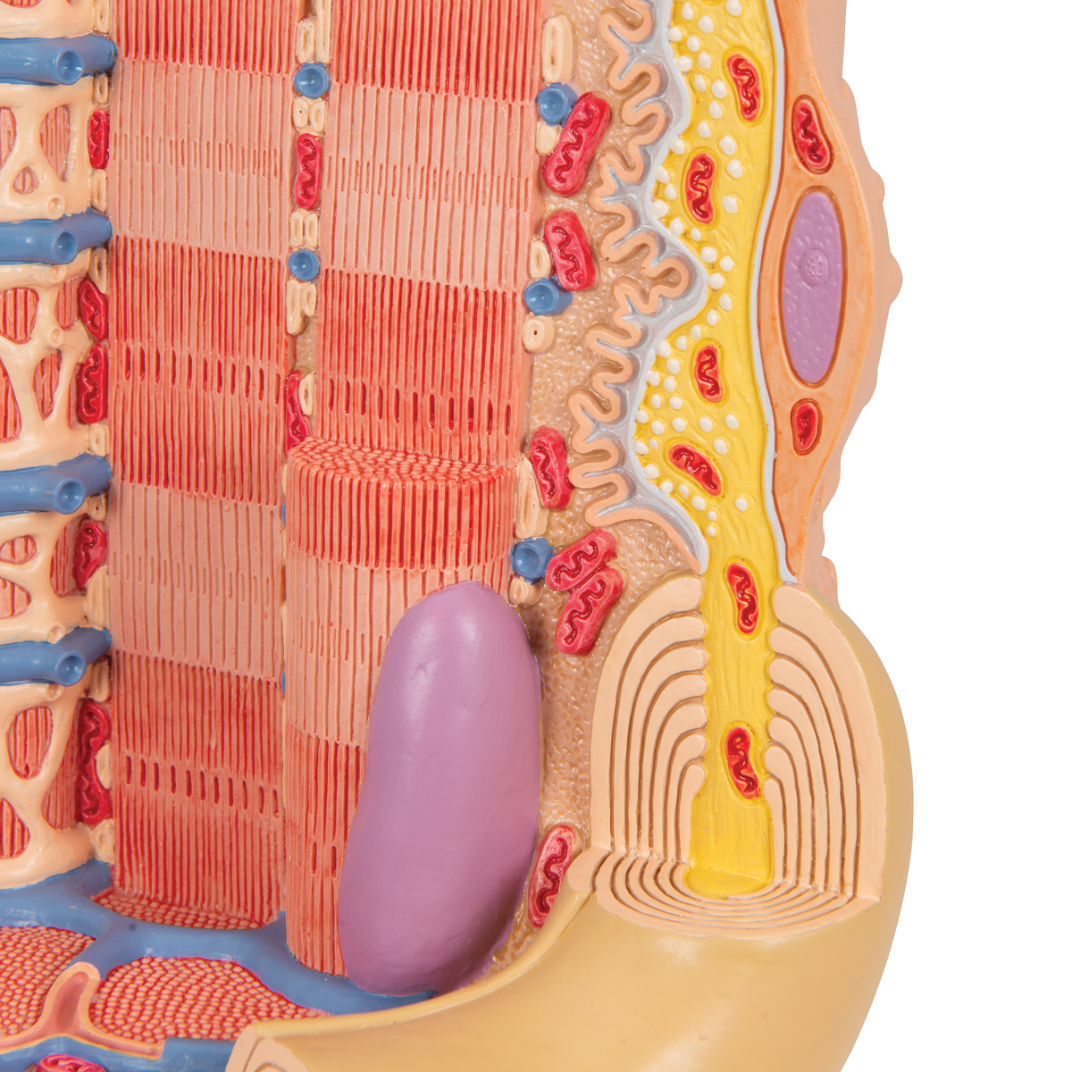
Sarcomere Model | Muscle Fiber Model | Skeletal Muscle | MICROanatomy ... This micro-anatomy model magnifies the anatomy of the human muscle fiber approximately 10,000 times. This muscle model illustrates a section of a skeletal muscle fiber and its neuromuscular end plate. The muscle fiber is the basic element of the diagonally striped skeletal muscle. You've never seen a muscle fiber in this way!
Systems Biology of Skeletal Muscle: Fiber Type as an Organizing Principle There are two isoforms of TnC: the cTnC ("c" for cardiac) isoform is expressed in cardiac and slow skeletal muscle fibers, whereas sTnC ("s" for skeletal) is expressed only in fast muscle fibers.
Nervous system Skeletal Muscle Fiber with Motor End-plate Human Anatomy ... High quality Nervous system Skeletal Muscle Fiber with Motor End-plate Human Anatomy Model for medical education from China, China's leading human anatomy simulation product, with strict quality control anatomy lab models factories, producing high quality anatomy lab models products.
Sarcomere Model | Muscle Fiber Model | Skeletal Muscle | MICROanatomy ... This micro-anatomy model magnifies the anatomy of the human muscle fiber approximately 10,000 times. This muscle model illustrates a section of a skeletal muscle fiber and its neuromuscular end plate. The muscle fiber is the basic element of the diagonally striped skeletal muscle. You've never seen a muscle fiber in this way!
Skeletal Muscle Anatomy Model Muscle and motion. Skeletal Muscle Anatomy Model. human bones diagram | Anatomy System - Human Body Anatomy diagram and we have 8 Pics about human bones diagram | Anatomy System - Human Body Anatomy diagram and like MUSCULAR SYSTEM ANATOMY:Muscle fiber with neuromuscular junction model, Muscle Fiber with motor end plate model Physiology Anatomy ...
MUSCULAR SYSTEM ANATOMY:Muscle fiber with neuromuscular ... Mar 28, 2012 ... Short video of a muscle fiber with the neuromuscular junction in ... MUSCULAR SYSTEM ANATOMY:Muscle fiber with neuromuscular junction model ...
skeletal anatomy labeled - Microsoft Muscle skeletal fiber contraction. Visible interactive iguana Neuromuscular Junction Model - YouTube. 10 Images about Neuromuscular Junction Model - YouTube : Image result for skeletal system labeled | Skeletal system worksheet, Human male skeleton - Stock Image - C024/9740 - Science Photo Library and also Slide 34 - Skeletal Muscle - YouTube.
Smooth Muscle | Boundless Anatomy and Physiology | | Course Hero These myoblasts asre located to the periphery of the myocyte and flattened so as not to impact myocyte contraction. Myocyte: Skeletal muscle cell: A skeletal muscle cell is surrounded by a plasma membrane called the sarcolemma with a cytoplasm called the sarcoplasm. A muscle fiber is composed of many myofibrils, packaged into orderly units.
Skeletal muscle fiber model Quiz - PurposeGames.com About this Quiz This is an online quiz called Skeletal muscle fiber model There is a printable worksheet available for download here so you can take the quiz with pen and paper. This quiz has tags. Click on the tags below to find other quizzes on the same subject. Anatomy Muscles muscle fiber Your Skills & Rank Total Points 0 Get started!
› science › articleMitochondrial dynamics maintain muscle stem cell regenerative ... Sep 01, 2022 · For muscle regeneration experiments, mice were anesthetized with isoflurane inhalation. Regeneration of skeletal muscle was induced by intramuscular injection of cardiotoxin (CTX, Latoxan) in the tibialis anterior (TA), gastrocnemius, and quadriceps muscle of the mice as previously described (Suelves et al., 2007). At the indicated times after ...
en.wikipedia.org › wiki › MyofibrilMyofibril - Wikipedia A myofibril (also known as a muscle fibril or sarcostyle) is a basic rod-like organelle of a muscle cell. Skeletal muscles are composed of long, tubular cells known as muscle fibers, and these cells contain many chains of myofibrils. Each myofibril has a diameter of 1–2 micrometres.
Skeletal Muscle Fiber Structure and Function - Open Textbooks for Hong Kong Figure 16.18 A skeletal muscle fiber is surrounded by a plasma membrane called the sarcolemma, with a cytoplasm called the sarcoplasm. A muscle fiber is composed of many fibrils packaged into orderly units. The orderly arrangement of the proteins in each unit, shown as red and blue lines, gives the cell its striated appearance.
Page: Journal of Hand Surgery Jul 13, 2022 · The Journal of Hand Surgery publishes original, peer-reviewed articles related to the pathophysiology, diagnosis, and treatment of diseases and conditions of the upper extremity; these include both clinical and basic science studies, along with case reports.
SKELETAL MUSCLE ORGANIZATION With your newly labeled image in hand, read through the following paragraphs. ... Each skeletal muscle cell, also called a muscle fiber, develops as many ...
Muscle Fiber Model: Motor Neuron, Myeline Sheath ... - Pinterest Dec 20, 2013 - Muscle Fiber Model: Motor Neuron, Myeline Sheath, Node of Ranvier, ... Functional anatomy of the skeletal muscle and muscle fibers.
muscle fiber anatomy diagram Muscle Fiber Model Labeled | Muscle Anatomy, Skeletal Muscle Anatomy . muscle cell anatomy skeletal fiber labeled hypertrophy body. Skeletal Muscle Anatomy Quiz - Quizizz quizizz.com. skeletal otot rangka lurik tubuh quizizz biologi fungsinya jantung soal beserta gambarnya kimia pngio jaringan usaha321 materikimia pngsumo
› doi › 10Semirational bioengineering of AAV vectors with increased ... Sep 21, 2022 · In a mouse model of X-linked myotubular myopathy, the best vectors—AAVMYO2 and AAVMYO3—prolonged survival, corrected growth, restored strength, and ameliorated muscle fiber size and centronucleation. In a mouse model of Duchenne muscular dystrophy, our lead capsid induced robust microdystrophin expression and improved muscle function.
2-Deoxy-D-glucose Alleviates Cancer Cachexia-Induced Muscle … Web25/09/2022 · Cachexia is characterized by progressive weight loss accompanied by the loss of specific skeletal muscle and adipose tissue. Increased lactate production, either due to the Warburg effect from tumors or accelerated glycolysis effects from cachectic muscle, is the most dangerous factor for cancer cachexia. This study aimed to explore the efficiency …
Semirational bioengineering of AAV vectors with increased … Web21/09/2022 · In a mouse model of X-linked myotubular myopathy, the best vectors—AAVMYO2 and AAVMYO3—prolonged survival, corrected growth, restored strength, and ameliorated muscle fiber size and centronucleation. In a mouse model of Duchenne muscular dystrophy, our lead capsid induced robust microdystrophin …
Home Page: Journal of Hand Surgery Web13/07/2022 · The Journal of Hand Surgery publishes original, peer-reviewed articles related to the pathophysiology, diagnosis, and treatment of diseases and conditions of the upper extremity; these include both clinical and basic science studies, along with case reports.Special features include Review Articles (including Current Concepts and The …
10.2 Skeletal Muscle – Anatomy & Physiology Describe the structure and function of skeletal muscle fibers ... This process is known as the sliding filament model of muscle contraction (Figure 10.2.4).
Mitochondrial dynamics maintain muscle stem cell regenerative ... Web01/09/2022 · Skeletal muscle satellite cells ... Using a genetic model ablating Drp1 in SCs, we show that mitochondrial dynamics, and in particular mitochondrial fission, are required for efficient expansion of SCs during muscle regeneration, by dictating mitochondrial metabolism and proteostatic fitness. We further reveal that reduced mitochondrial fission …
10.2 Skeletal Muscle - Anatomy and Physiology 2e | OpenStax Apr 20, 2022 ... Because skeletal muscle cells are long and cylindrical, they are commonly referred to as muscle fibers. Skeletal muscle fibers can be quite ...
Altay Skeletal Muscle Fiber Model | Carolina.com $291.75 Quantity (in stock) add to wishlist Description Altay®. 10,000× life size. Microstructure of a skeletal muscle fiber is represented in great detail; includes the neuromuscular junction, sarcoplasmic reticulum, T tubules, and myofibrils. Mounted on reinforced polymer base. Size, 25 × 23 × 23 cm. Specifications Return Policy:
tech.msu.edu › about › guidelines-policiesAndrew File System Retirement - Technology at MSU Andrew File System (AFS) ended service on January 1, 2021. AFS was a file system and sharing platform that allowed users to access and distribute stored content. AFS was available at afs.msu.edu an…
muscle fiber model labeling Diagram | Quizlet Only $35.99/year muscle fiber model labeling How do you want to study today? Flashcards Review terms and definitions Learn Focus your studying with a path Test Take a practice test Match Get faster at matching terms Created by crlavenuePLUS Terms in this set (7) transverse tubule sarcoplasmic reticulum triad sarcolemma myofibril
Correctly Label The Following Parts Of A Skeletal Muscle Fiber A skeletal muscle fiber is surrounded by a plasma membrane, which contains the sarcoplasm of the muscle cell. It is composed of many fibrils. These fibrils give the muscle fiber its striated appearance. The myofilaments are arranged sequentially in the sarcomere, which gives the fiber its striated look.














:background_color(FFFFFF):format(jpeg)/images/article/en/the-neuromuscular-junction-structure-and-function/y6U7kRYvPqiGcdT2v0SFPQ_0KDfoRV908RqFJjJfQUAg_132Neuromuscular_junction_magnified.png)


Komentar
Posting Komentar