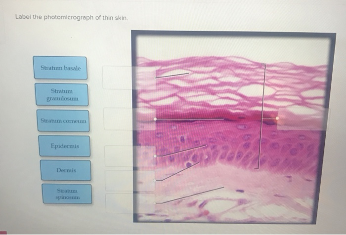
40 label the photomicrograph of thin skin
C ezto.mheducation.com/hm.tpx 23. Label the photomicrograph of thin ... Label the photomicrograph of thin skin. Hair Sebaceous gland Dermis Hair Follicle Epidermis Duct of sebaceous gland... Questions & Answers. Accounting. Financial Accounting; Cost Management; Managerial Accounting; Advanced Accounting; Auditing; Accounting - Others; Accounting Concepts and Principles; Label The Photomicrograph - Mr. Hill's Biology Blog: Our cells "inner skin" Label the photomicrograph of thick skin. Use a label line and the letter p for each section. Monocyte, erythrocyte, lymphocyte, neutrophil, basophil, eosinophil. Schematically sketch and label the resulting microstructure. Place the following layers in order from superficial to deep.
photomicrograph of thick skin Diagram | Quizlet Start studying photomicrograph of thick skin. Learn vocabulary, terms, and more with flashcards, games, and other study tools.

Label the photomicrograph of thin skin
Label The Photomicrograph Of Thick Skin - Faktor yang Label the photomicrograph of thick skin. 1 answer to label the photomicrograph of thin skin. The epidermis of thick skin has five layers: Hypodermis label the layers of the epidermis in thick skin in figure 7.2. A few layers of cells that are . Apocrine sweat gland label the photomicrograph in figure 7.4. Label the photomicrograph of thick skin. Anatomy and Physiology Homework Chapter 6 Flashcards | Quizlet Study with Quizlet and memorize flashcards containing terms like Label the parts of the skin and subcutaneous tissue. -Blood Capillaries -Piloerector muscle -Dermal papilla -Hair bulb -Sensory nerve fibers -Tactile corpuscle -Hair follicle -Sebaceous gland, Label the parts of the skin and subcutaneous tissue. -Hypodermis -Sweat pores -Dermis -Hairs -Cutaneous blood vessels -Epidermis -Sweat ... Label The Photomicrograph Of Thick Skin Quizlet : Organs And Structures ... Label the photomicrograph of thick skin. Label the structures of the skin and subcutaneous tissues. It consists of three main la. Epidermis, stratum corneum, stratum lucidum, stratum granulosum, stratum spinosum, stratum basale, dermis. Which of the following are characteristics of thick skin?
Label the photomicrograph of thin skin. Sebaceous Gland Label The Photomicrograph Of Thin Skin - Blogger Label the photomicrograph of thin skin. The skin and its associated structures, hair, sweat glands and nails make up . The ducts are lined by stratified (2 layers) cuboidal epithelium. Label the photomicrograph of thin skin 3 10 points duct of sebaceous gland references epidermis hair follicle hair dermis sebaceous . A&P 1 Exercise_7 Activity 1 & 2 & RYK and UYK.docx - LAB... Apocrine sweat Gland Label the photomicrograph in Figure 7.4. 1. Sebaceous glands 2. Hair follicle 3. Hair root 4. Hair bulb 5. Papilla of hair ... Translucent layer found in thick skin, absent in thin skin. Stratum Spinosum 6. Appears to have thorn-like projections in prepared slides. Reticular Region 7. PDF The Integumentary System - Holly H. Nash-Rule, PhD Label the skin structures and areas indicated in the accompanying diagram of thin skin. Then, complete the statements that follow. a. Lamellar granules contain glycolipids that prevent water loss from the skin. b. Fibers in the dermis are produced by fibroblasts . photomicrographs of thin skin Flashcards | Quizlet photomicrographs of thin skin. Term. 1 / 4. stratum corneum. Click the card to flip 👆. Definition. 1 / 4. ... Click the card to flip 👆.
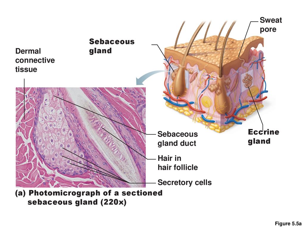
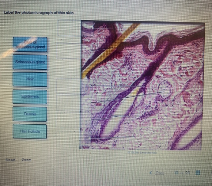
unit 4 lab.docx - LAB Unit 4 EXERCISE 7: The Integumentary... FIGURE 7.4: Diagram of the skin and accessory structures. • apocrine (AP-oh-krin) sweat gland • arrector pili (PIE-lee) muscle • eccrine (EK-rin) sweat gland • hair bulb • hair follicle • hair root • hair shaft • papilla (puh-PILL-uh) of hair • sebaceous (se-BAY-shus) gland 1. Hair shaft 2. Hair root 3. Sebaceous glands 4. Arrector pili muscle 5. Photomicrograph of Thick Skin Quiz - PurposeGames.com This is an online quiz called Photomicrograph of Thick Skin There is a printable worksheet available for download here so you can take the quiz with pen and paper. Your Skills & Rank Total Points 0 Get started! Today's Rank -- 0 Today 's Points One of us! Game Points 6 You need to get 100% to score the 6 points available Actions Add to Playlist Question : Label the photomicrograph of thin skin. Dermis Duct of ... Expert Answer. 100% (37 ratings) A …. View the full answer. Transcribed image text: Label the photomicrograph of thin skin. Dermis Duct of sebaceous gland Hair Follicle Sebaceous gland Hair Epidermis. Question : Question 31 points Label the photomicrograph of thin skin ... 31 points Label the photomicrograph of thin skin. Hair Follicle Hair Dermis Sebaceous gland Duct of sebaceous gland Reset zoom Solution 5 (1 Ratings ) Solved Biology 2 Years Ago 77 Views This Question has Been Answered! View Solution Question 31) True breeding white flowering plants are crossed to true breeding red flowering plants.
PDF Name the Condition Name the 4 layers of thin skin in both the cartoon and the photomicrograph. Name the 4 layers of thin skin in both the cartoon and the photomicrograph. •Name the Layers of skin and label the dermal papilla and dermis •Name the Layers of skin and label the dermal papilla Quiz #3 Study Guide Flashcards | Quizlet Label the photomicrograph of thin skin. Organize the following layers of the epidermis from superficial to deep. Categorize the appropriate structures or descriptions with the appropriate layer of skin that is highlighted in blue. Words can be used more than once. Top section 1. Composed of 5 layers 2. Is avascular 3. Composed of keratinocytes 4. Most superficial layer … Figure 7.1: Photomicrograph of Skin Diagram | Quizlet Start studying Figure 7.1: Photomicrograph of Skin. Learn vocabulary, terms, and more with flashcards, games, and other study tools. anatomy lab, exam 3, lab 9, Spinal Nerves, Integument, and ... - Quizlet Label the photomicrograph of thin skin. stratum corneum stratum granulosum stratum spinosum stratum basale dermis epidermis hypodermis the layer of skin beneath the dermis, which serves as a storage repository for fat Name the yellow highlighted structures that pass through the intervertebral foramina. spinal nerves
SciELO - Brasil - Autopsy findings in a patient with primary ...
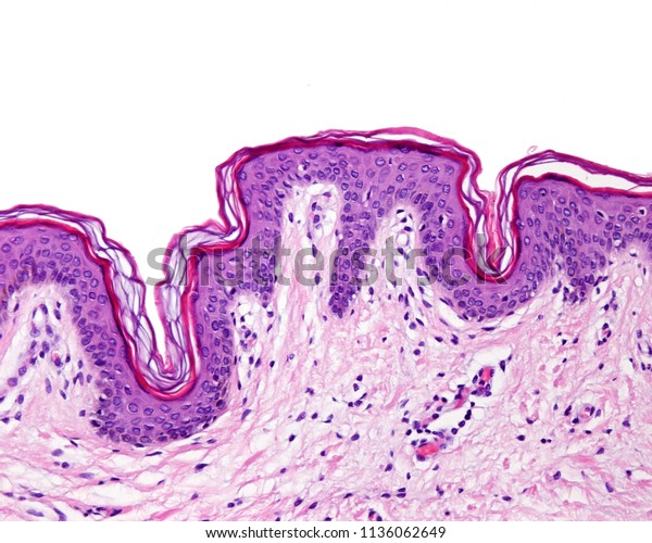
Layers of the Skin | Anatomy and Physiology I | | Course Hero The cells in all of the layers except the stratum basale are called keratinocytes. A keratinocyte is a cell that manufactures and stores the protein keratin. Keratin is an intracellular fibrous protein that gives hair, nails, and skin their hardness and water-resistant properties.The keratinocytes in the stratum corneum are dead and regularly slough away, being replaced by cells from the ...
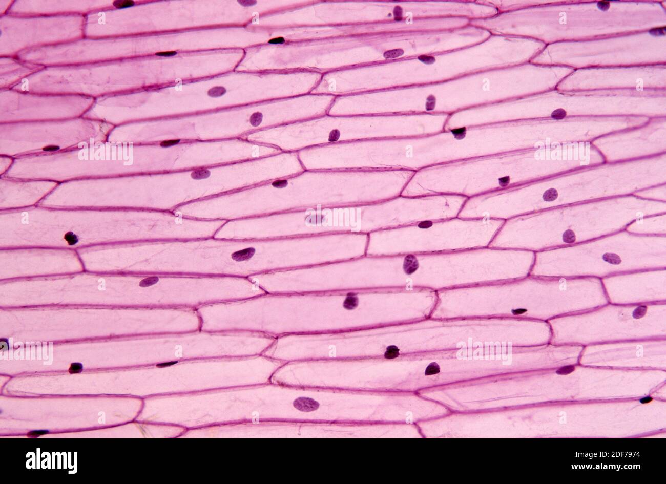
Epidermis of onion (Allium cepa) with cells, nucleus and ...
Label The Photomicrograph Of Thick Skin. - Martina Eisenhower Take several photomicrographs of thin skin at this magnification. 1 answer to label the photomicrograph of thin skin. Stratum basale, stratum spinosum, stratum granulosum, stratum lucidum, and stratum . It has a fifth layer,. Learn more about skin discoloration treatments in this quick guide.

Time-Dependent Effect of Oral Morphine Consumption on the ...
Label the photomicrograph in Figure 7.4. Examine a slide of hairy skin ... Label The Photomicrograph Of The Skin And Its Accessory Structures. Sebaceous Gland Duct Of Sebaceous Gland Epidermis Hair Follicle ... Activity 4 Differentiating Sebaceous and Sweat Glands Microscopically Using the slide thin skin with hairs and the photomicrographs of cutaneous glands (Figure 7.6) as a guide, identify sebaceous and eccrine ...

Changes in Dermal Thickness in Biopsy Study of Histologic ...
Leaf micrograph section hi-res stock photography and images - Alamy RM JTREDJ - Snapdragon rust, Puccinia antirrhini fungus on antirrhinum leaf, darkfield photomicrograph, TS. RF D6509D - Tea leaf, light micrograph. RM M01FYF - Bright field light micrograph of a cotton plant leaf, pictured area is 1.7mm wide. RF 2GAFY36 - Cross section of a leaf, light micrograph.

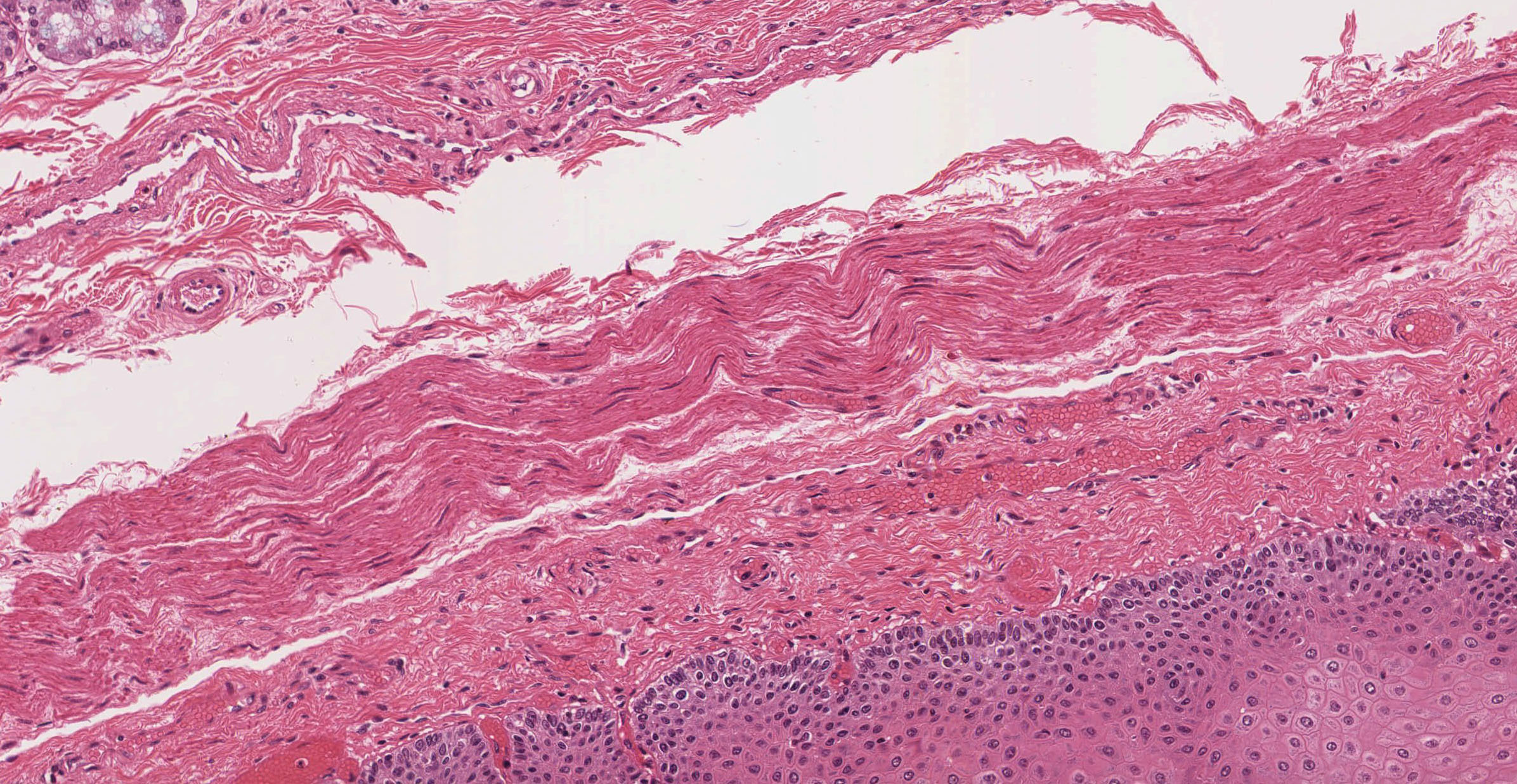
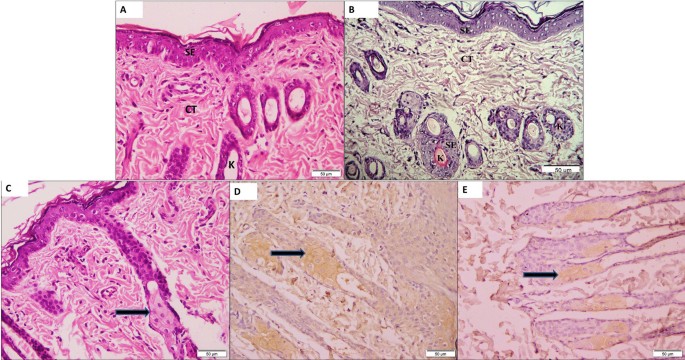
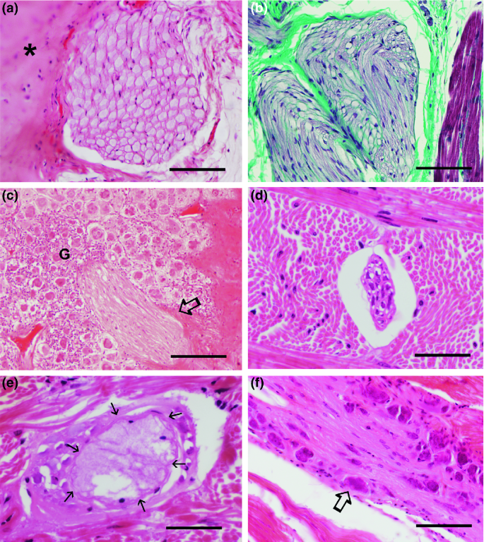
a): A photomicrograph of the section of thin skin tissue from ...
Label The Photomicrograph Of Thick Skin / Solved Label The ... - Blogger The epidermis of thick skin has five layers: Thick skin · stratum basale (also known as s. Label the photomicrograph of thick skin. It has a fifth layer,. Start studying photomicrograph of the epidermal layer in thick skin. The outer layer of cells in this micrograph is the thinnest layer and. A few layers of cells that are .
Epidermis Thin Skin Can Be Identified Foto Stok 1136062649 ...
Cambridge International AS & A Level Biology Coursebook … A photomicrograph is a photograph of a specimen as seen with a light microscope. Figure 1.6 shows some human cells. Figure 1.7 shows a plant cell taken from a leaf. Both figures show cells magnified 400 times, which is equivalent to using the high-power objective lens on a light microscope. See also Figures 1.8a and 1.8b for labelled drawings of these figures.

Pin by nico x. on Anatomy | Games, Tetris, Anatomy
Cambridge International AS and A Level Biology ... - Academia.edu BIO1: Maintaining a Balance 1. Most organisms are active in a limited temperature range IDENTIFY THE ROLE OF ENZYMES IN METABOLISM, DESCRIBE THEIR CHEMICAL COMPOSITION AND USE A SIMPLE MODEL TO DESCRIBE …
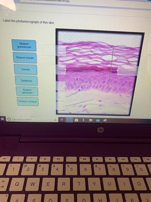
Solved met Label the photomicrograph of thin skin Stratum ...
(Solved) - Label the photomicrograph of thin skin. O ... - Transtutors 1 Answer to Label the photomicrograph ...
Pharynx, Esophagus, and Stomach | histology
(Solved) - Label The Photomicrograph Of The Skin And Its Accessory ... Sebaceous gill: The sebum ...

Layers of the Skin | Anatomy and Physiology I
Label The Photomicrograph Of Thick Skin Quizlet : Organs And Structures ... Label the photomicrograph of thick skin. Label the structures of the skin and subcutaneous tissues. It consists of three main la. Epidermis, stratum corneum, stratum lucidum, stratum granulosum, stratum spinosum, stratum basale, dermis. Which of the following are characteristics of thick skin?
Implanted subcutaneous versus intraperitoneal bioscaffold ...
Anatomy and Physiology Homework Chapter 6 Flashcards | Quizlet Study with Quizlet and memorize flashcards containing terms like Label the parts of the skin and subcutaneous tissue. -Blood Capillaries -Piloerector muscle -Dermal papilla -Hair bulb -Sensory nerve fibers -Tactile corpuscle -Hair follicle -Sebaceous gland, Label the parts of the skin and subcutaneous tissue. -Hypodermis -Sweat pores -Dermis -Hairs -Cutaneous blood vessels -Epidermis -Sweat ...
Solved Label these structures located in axillary skin. Hair ...
Label The Photomicrograph Of Thick Skin - Faktor yang Label the photomicrograph of thick skin. 1 answer to label the photomicrograph of thin skin. The epidermis of thick skin has five layers: Hypodermis label the layers of the epidermis in thick skin in figure 7.2. A few layers of cells that are . Apocrine sweat gland label the photomicrograph in figure 7.4. Label the photomicrograph of thick skin.
Functional Histology: The Tissues of Common Coleoid ...

Layers of the Skin | Anatomy and Physiology I

SciELO - Brasil - Oro-facial-digital syndrome type I: a case ...

Photomicrograph of Thin Skin Quiz

Photomicrographs of skin (Thick skin) Diagram | Quizlet
Integumentary System Overview
Integument System. - ppt download

a): A photomicrograph of the section of thin skin tissue from ...

Calcium-sensing receptor deletion in the mouse esophagus ...
Histology of major organ systems of Nothobranchius fishes ...
Handout: The Integumentary System Anatomy & Physiology I ...
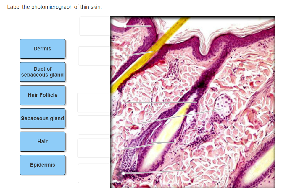
Solved Label the photomicrograph of thin skin. Dermis Duct ...
:max_bytes(150000):strip_icc()/5324695-GettyImages-139812232-75c6744d0b2246fba58223c0eb784c73.jpg)
Hypodermis (Subcutaneous Tissue): Anatomy and Function
Solved Label the photomicrograph of thin skin. deous gland ...
Hair follicle in skin hi-res stock photography and images - Alamy

Supraclavicular extra-renal angiomyolipoma: a challenging ...
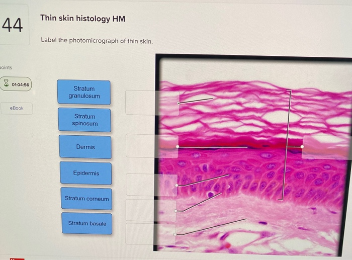
Solved Thin skin histology HM 44 Label the photomicrograph ...

SciELO - Brasil - Macroscopic and microscopic morphology of ...
The Role of Hesperidin on Healing an Incised Wound in an ...

Pathogenesis of Aeromonas caviae in Clarias magur - ScienceDirect
Histology Of Skin | Faculty of Medicine

a): A photomicrograph of the section of thin skin tissue from ...

Osteochondrolipoma of the Hand - Journal of Hand Surgery

a) A photomicrograph of the section of thin skin tissue from ...
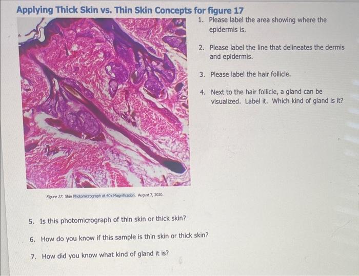
Applying Thick Skin vs. Thin Skin Concepts for figure | Chegg.com
Integumentary System | histology

Lap Practical #1 EC Flashcards | Quizlet

The microscopic image of the time-dependent effect of oral ...


Komentar
Posting Komentar